

Analysis of the collected sample should be performed by an experienced Microbiolo- gist, Aerobiologist, Mycologist or Environmental Microscopist. Counting and quanti- fication of sample components is conducted by counting calibrated cross-sections of the deposited sample trace. The number and type of particles counted per cubic meter of air is calculated based on the length of the deposition trace, length of trace actually examined, volume of air collected, and number of particles counted.
The Air-O-Cell particle deposition area at a flow rate of 15 lpm is approximately 1.1 mm wide by 14.5 mm long yielding an approximate area of 15.95 mm2. The width of the deposition trace will vary slightly with flow rate and media thickness, and will vary slightly in particle density from the middle to outer edges of deposi- tion. For this reason, using the deposition trace area is not recommended for direct calculation of particle concentrations. The recommended procedure for calculating particle concentrations is based on using the Air-O-Cell trace length and microscope field diameter, and will be discussed below. One field of view counted is defined as the calibrated diameter of the microscope field of view (in mm) covering one cross- sectional pass or “traverse” across the sample deposition trace. A typical sample preparation and microscopic counting procedure is illustrated in Figure 2. 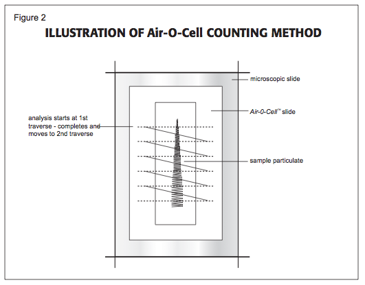
The calculation of particle concentration per cubic meter of air can be performed by using the following equations.
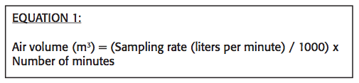
First, determine the actual air volume collected in cubic meters (m3) by following the calculation given in Equation 1.
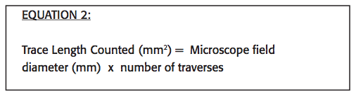
Second, determine the length of sample trace counted based on the microscope field of view and number of fields of view counted. Accurately calibrate and measure the diameter of the microscope field of view using a stage micrometer slide. Remember, each microscope is different, and each different combination of ocular and objec- tive lens must be calibrated separately. Stated lens magnifications are rarely precise. The microscopist should then record the number of complete traverses examined across the width of the deposition trace and use the formula given in Equation 2 to calculate the actual length of the deposition trace examined.
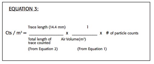
The concentrations of particles (cts/m3) can then be determined by using Equation 3.
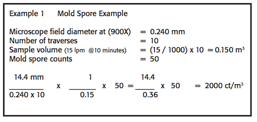
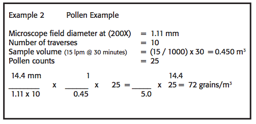
Two example calculations for mold spores and pollen grains are given below:
Comments